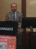
Prof. Orestis Ioannidis
General Hospital ‘G. Papanikolaou”, Greece
Title: Open abdomen and negative pressure wound therapy for acute peritonitis especially in the presence of anastomoses and ostomies
Abstract:
Acute peritonitis is a relatively
common intra-abdominal infection that a general surgeon will have to manage
many times in his surgical carrier. Usually it is a secondary peritonitis
caused either by direct peritoneal invasion from an inflamed infected viscera
or by gastrointestinal tract integrity loss. The mainstay of treatment is
source control of the infection which is in most cases surgical. In the
physiologically deranged patient there is indication for source control surgery
in order to restore the patient’s physiology and not the patient anatomy
utilizing a step approach and allowing the patient to resuscitate in the
intensive care unit. In such cases there is a clear indication for relaparotomy
and the most common strategy applied is open abdomen. In the open abdomen
technique the fascial edges are not approximated and a temporarily closure
technique is used. In such cases the negative pressure wound therapy seems to
be the most favourable technique, as especially in combination with fascial
traction either by sutures or by mesh gives the best results regarding delayed
definite fascial closure, and morbidity and mortality. In our surgical practice
we utilize in most cases the use of negative pressure wound therapy with a
temporary mesh placement. In the initial laparotomy the mesh is placed to
approximate the fascial edges as much as possible without whoever causing
abdominal hypertension and in every relaparotomy the mesh is divided in the
middle and, after the end of the relaparotomy and dressing change, is
approximated as much as possible in order for the fascial edges to be further
approximated. In every relaparotomy the mesh is further reduced to finally
allow definite closure of the aponeurosis. In the presence of ostomies the
negative pressure wound therapy can be applied as usual taking care just to
place the dressing around the stoma and the negative pressure can be the
standard of -125 mmHg. However, in the presence of anastomosis the available
date are scarce and the possible strategies are to differ the anastomosis for the
relaparotomy with definitive closure and no further need of negative pressure
wound therapy, to low the pressure to -25 mmHg in order to protect the
anastomosis and to place the anastomosis with omentum in order to avoid direct
contact to the dressing. The objective should be early closure, within 7 days,
of the open abdomen to reduce mortality and complications.
Use of
indocyanine green fluorescence imaging in the extrahepatic biliary tract
surgery
Cholelithiasis presents in approximately 20 % of the total population, ranging between 10% and 30 %. It presents one of the most common causes for non malignant surgical treatment. The cornerstone therapy is laparoscopic cholecystectomy, urgent of elective. Laparoscopic cholecystectomy is nowadays the gold standard surgical treatment method, however bile duct injury occurred to as high as 0.4-3% of all laparoscopic cholecystectomies. The percentage has decreased significantly to 0.26-0.7% because of increased surgical experience and advances in laparoscopic imaging the past decade which have brought to light new achievements and new methods for better intraoperative visualization such as HD and 3D imaging system. However, bile duct injury remains a significant issue and indocyanine green fluorescence imaging, mainly cholangiography but also angiography, can further enhance the safety of laparoscopic cholecystectomy as it allows the earlier recognition of the cystic and common bile duct, even in several times before dissecting the Callot triangle. Fluorescence cholangiography could be an ideal method in order to improve bile tree anatomy identification and enhance prevention of iatrogenic injuries during laparoscopic cholecystectomies and also it could be helpful in young surgeons training because it provides enhanced intraoperative safety, but however this method does not replace CVS. Finally, our ongoing current study results comparing intravenous to direct administration of ICG in the gallbladder will be presented.
Biography:
Dr.
Ioannidis is currently an Assistant Professor of Surgery in the Medical School
of Aristotle University of Thessaloniki. He studied medicine in the Aristotle
University of Thessaloniki and graduated at 2005. He received his MSC in
“Medical Research Methodology” in 2008 from Aristotle University of
Thessaloniki and in “Surgery of Liver, Biliary Tree and Pancreas” from the
Democritus University of Thrace in 2016. He received his PhD degree in 2014
from the Aristotle University of Thessaloniki as valedictorian for his thesis
“The effect of combined administration of omega-3 and omega-6 fatty acids in
ulcerative colitis. Experimental study in rats.” He is a General Surgeon with
special interest in laparoscopic surgery and surgical oncology and also in
surgical infections, acute care surgery, nutrition and ERAS and vascular
access. He has received fellowships for EAES, ESSO, EPC, ESCP and ACS and has
published more than 180 articles with more than 3000 citations and an H-index
of 28